43 microscope with labels
Microscope Types (with labeled diagrams) and Functions Simple microscope labeled diagram Simple microscope functions It is used in industrial applications like: Watchmakers to assemble watches Cloth industry to count the number of threads or fibers in a cloth Jewelers to examine the finer parts of jewelry Miniature artists to examine and build their work Also used to inspect finer details on products › cells › bactcellInteractive Bacteria Cell Model - CELLS alive Ribosomes: Ribosomes give the cytoplasm of bacteria a granular appearance in electron micrographs.Though smaller than the ribosomes in eukaryotic cells, these inclusions have a similar function in translating the genetic message in messenger RNA into the production of peptide sequences (proteins).
5 Types of Microscopes with Definitions, Principle, Uses, Labeled Diagrams 5 Types of Microscopes with Definitions, Principle, Uses, Labeled Diagrams March 1, 2022 by Sagar Aryal Subscribe us to receive latest notes. 5 Types of Microscopes Bright-Field or Light Microscope Dark Field Microscope Phase Contrast Microscope Fluorescence Microscope Electron Microscope Principle of Transmission Electron Microscope (TEM)

Microscope with labels
Microscope, Microscope Parts, Labeled Diagram, and Functions Microscope, Microscope Parts, Labeled Diagram, and Functions What is Microscope? A microscope is a laboratory instrument used to examine objects that are too small to be seen by the naked eye. It is derived from Ancient Greek words and composed of mikrós, "small" and skopeîn,"to look" or "see". › internet › portalCanon U.S.A., Inc. | EOS Utility EOS Utility is an application that brings together functions to communicate with the camera. These functions include downloading and displaying images, remote shooting, and camera control for each setting. Histology and Microscope Slide Labels Microscope Slide Labels. These specialty Microscope Slide Labels and matching End Labels are available in standard (thin) or pathology (tissue high) thickness, and square or round corner (RC). Permanent adhesive holds labels in place during use and long-term storage. Sheet Form Size is 5¼" x 8". Prices are per thousand labels. Slide Label
Microscope with labels. Binocular Microscope Anatomy - Parts and Functions with a Labeled ... Mechanical stages of the microscope, Frame or arm of a microscope, Objective lens (10X, 20X, 40X, and 100X), The nose piece of a microscope, Head part of the microscope, Ocular lens or eyepiece of the microscope, Diopter adjustment of the eyepiece All of these parts are identified in a light microscope labeled diagram. A Study of the Microscope and its Functions With a Labeled Diagram These labeled microscope diagrams and the functions of its various parts, attempt to simplify the microscope for you. However, as the saying goes, 'practice makes perfect', here is a blank compound microscope diagram and blank electron microscope diagram to label. Download the diagrams and practice labeling the different parts of these ... Compound Microscope Parts - Labeled Diagram and their Functions - Rs ... The eyepiece (or ocular lens) is the lens part at the top of a microscope that the viewer looks through. The standard eyepiece has a magnification of 10x. You may exchange with an optional eyepiece ranging from 5x - 30x. [In this figure] The structure inside an eyepiece. The current design of the eyepiece is no longer a single convex lens. Compound Microscope Parts, Functions, and Labeled Diagram Compound Microscope Definitions for Labels. Eyepiece (ocular lens) with or without Pointer: The part that is looked through at the top of the compound microscope. Eyepieces typically have a magnification between 5x & 30x. Monocular or Binocular Head: Structural support that holds & connects the eyepieces to the objective lenses.
Microscope With Labeled Parts And Functions Microscope With Labeled Parts and Functions Written by Editor in General Biology Microscope is a revolutionized scientific instrument which is used in research laboratories to examine the small objects that are not clearly visible and can't be seen by the naked eye. proscitech.com.auProSciTech Laboratory supplies and Lab equipment for Histology, Pathology, Light Microscopy, Electron Microscopy and specialist researchers. Parts of the Microscope with Labeling (also Free Printouts) Parts of the Microscope with Labeling (also Free Printouts) A microscope is one of the invaluable tools in the laboratory setting. It is used to observe things that cannot be seen by the naked eye. Table of Contents 1. Eyepiece 2. Body tube/Head 3. Turret/Nose piece 4. Objective lenses 5. Knobs (fine and coarse) 6. Stage and stage clips 7. Aperture Sperm Under Microscope with Labeled Diagram » AnatomyLearner >> The ... Sperm Under Microscope 400X Labeled Diagram Before that, you may also read the below-mentioned article to get a full idea of the structure of seminiferous tubules - Histological features of the seminiferous tubules with the labeled diagram Okay, first, let's see the different histological features of the seminiferous tubules of an animal.
Simple Microscope - Diagram (Parts labelled), Principle, Formula and Uses A simple microscope consists of Optical parts Mechanical parts Labeled Diagram of simple microscope parts Optical parts The optical parts of a simple microscope include Lens Mirror Eyepiece Lens A simple microscope uses biconvex lens to magnify the image of a specimen under focus. Parts of a microscope with functions and labeled diagram Microscopic illuminator - This is the microscopes light source, located at the base. It is used instead of a mirror. It captures light from an external source of a low voltage of about 100v. Condenser - These are lenses that are used to collect and focus light from the illuminator into the specimen. Microscope Labeled Pictures, Images and Stock Photos Browse 48 microscope labeled stock photos and images available, or start a new search to explore more stock photos and images. Newest results Fluorescent Imaging immunofluorescence of cancer cells growing... Plant Tissue Systems vector illustration. Labeled biology... Microscope diagram vector illustration. Labeled zoom instrument... Microscope Labeling - The Biology Corner I keep these instructions on the board for the week as a reminder: 1) Start with scanning (the shortest objective) and only use the COARSE knob . Once it is focused… 2) Switch to low power (medium) and only use the COARSE knob . You may need to recenter your slide. Once it is focused.. 3) Switch to high power (long objective).
Best Top Desktop Wallpapers: Electron microscope images
Microscope Parts, Function, & Labeled Diagram - slidingmotion Microscope parts labeled diagram gives us all the information about its parts and their position in the microscope. Microscope Parts Labeled Diagram The principle of the Microscope gives you an exact reason to use it. It works on the 3 principles. Magnification Resolving Power Numerical Aperture. Parts of Microscope Head Base Arm Eyepiece Lens

MICROBIOLOGY SLIDE SPECIMENS
Microscope Drawing And Label - Painting Valley Microscope Labeling ... 500x529 17 0 Label The Microscope... 270x350 14 0 Compound Light Micro... 630x380 8 1 Labeling The Parts O... 525x450 7 0 Compound Microscope ... 413x424 6 0 Microscope - Microsc... 236x262 4 1 Label Microscope Dia... 459x457 4 0 Microscope Parts Dia... 576x400 3 0 Section Cells View A... 512x346 2 1 Exe - Microscope Dra...
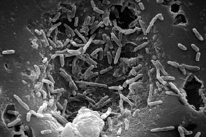
Mycobacterium leprae* - microbewiki
Label the microscope - Science Learning Hub Jun 8, 2018 — Use this interactive to identify and label the main parts of a microscope. Drag and drop the text labels onto the microscope diagram.Labels: DescriptionEye piece lens: The lens you look through – no...Light source: Sends light onto the specimen/slideCoarse focus adjustment: Moves the lens up or ...
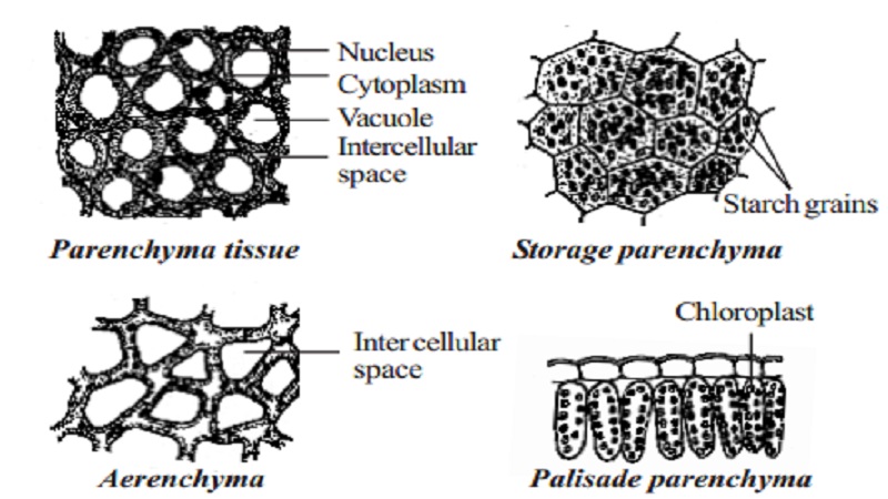
Permanent tissue: characteristics, types and functions - Online Biology ...
Microscope Parts and Functions Microscope Parts and Functions With Labeled Diagram and Functions How does a Compound Microscope Work?. Before exploring microscope parts and functions, you should probably understand that the compound light microscope is more complicated than just a microscope with more than one lens.. First, the purpose of a microscope is to magnify a small object or to magnify the fine details of a larger ...
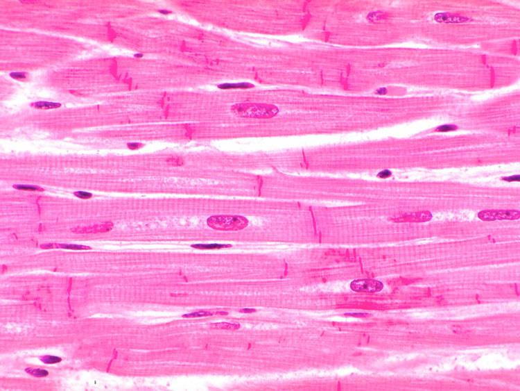
Tissues Flashcards | Easy Notecards
Microscope Labeling Game - PurposeGames.com About this Quiz. This is an online quiz called Microscope Labeling Game. There is a printable worksheet available for download here so you can take the quiz with pen and paper. This quiz has tags. Click on the tags below to find other quizzes on the same subject. Science.
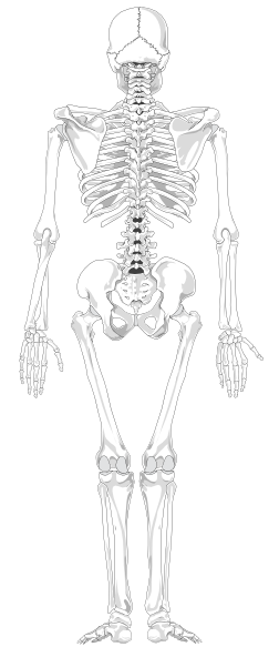
Human Skeleton Back No Text No Color Clip Art at Clker.com - vector ...
› products › confocal-microscopesOverview of Confocal Laser Scanning Microscopes - ZEISS 4D super-resolution imaging and simultaneous spectral detection of multiple weak labels up to 900 nm emission. LSM 900 with Airyscan 2 Your compact confocal for gentle multiplex imaging and smart analysis

Tongue Histology | Histology slides, Human anatomy and physiology ...
Microscope labels | Bill Todt Microscope labels. Unlabelled image: Lab Index: 101 Class Page: Identify the following parts of the microscope: Eyepieces Observation tube Nosepiece Objectives Stand Specimen holder Mechanical stage Stage controls Condenser Iris diaphragm Coarse focus Fine focus Base Rheostat Power switch.
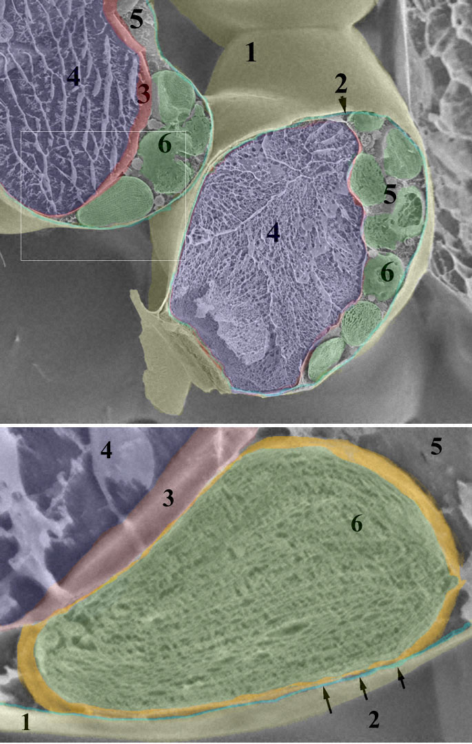
Leaf chloroplast
› watchEvery IB Biology drawing you NEED to know - YouTube PLEASE READ!This is every single drawing you need to know!Looking through the 2016 syllabus, this video covers 2 main types of statements- Draw- Annotate ( +...

Microscope Clip Art at Clker.com - vector clip art online, royalty free ...
Histology and Microscope Slide Labels Microscope Slide Labels. These specialty Microscope Slide Labels and matching End Labels are available in standard (thin) or pathology (tissue high) thickness, and square or round corner (RC). Permanent adhesive holds labels in place during use and long-term storage. Sheet Form Size is 5¼" x 8". Prices are per thousand labels. Slide Label
Post a Comment for "43 microscope with labels"